Drafting
Collaboration between cardiology and nuclear medicine services began more than 30 years ago. Thanks to the diagnostic techniques offered by nuclear medicine, cardiologists have been able to speed up and improve diagnosis, while providing greater comfort to their patients. Within the framework of Heart Month, specifically World Heart Day celebrated on September 29, Dr. Ricardo Ruano PerezChairman of the Nuclear Cardiology Working Group Spanish Society of Nuclear Medicine and Molecular Imaging (Semnem)It addresses the joint work undertaken by both specialties and its impact on patients.
Collaboration between nuclear medicine and cardiology began about 30 years ago
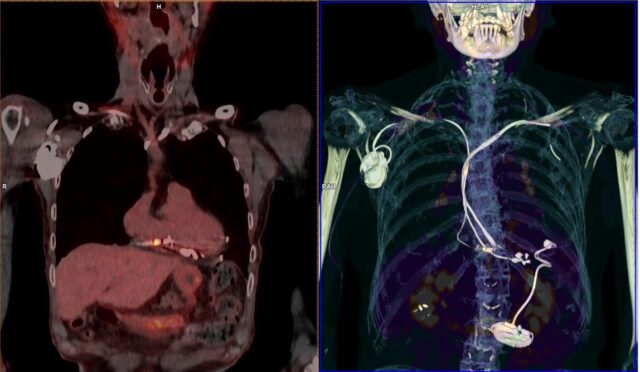
As Dr. Ruano explains, collaboration between both fields has been carried out for more than 30 years using single photon emission computed tomography (SPECT) in ischemic heart diseases. “Recently, positron emission tomography (PET) tests in the diagnosis of infection (endocarditis) have become the most common indication for cardiovascular disease; The radiopharmaceutical used is called FDG (fluordeoxyglucose) and has a high diagnostic capacity that facilitates the establishment of treatment.“, adds the specialist.
Digitization and artificial intelligence in diagnosis
Dr. Ruano highlights that “In the past two years, the latest equipment has been integrated, namely PET-CT digital tomography, which uses artificial intelligence tools and represents a significant improvement in diagnostic performance.“.”At the cardiovascular level, the improvement is noticeable with increased accuracy and diagnostic capacity, allowing studies to be performed in less time and thus improving patient comfort.says Dr. Ruano.
These developments also have an impact on diagnostic accuracy. “Technological development, with more precise and accurate diagnostic equipment, with improved software focused on process automation, ensures accuracy in diagnosis.“, points out Dr. Ruano.
Advances in diagnostic equipment make the diagnosis more accurate
Furthermore, Dr. Ruano points to this progress in digitizing, processing and storing heart images.It is a major advance that facilitates rapid diagnosis and thus allows choosing the best treatment for a heart patient.“.
The impact of diagnostic imaging in heart diseases
Within the framework of the 40th SEMNIM National Congress, a roundtable addressed the paradigm shift in diagnostic imaging equipment. And so, Dr. Virginia Alvarez Asiaen, Positron Emission Tomography (PET) scan, highlighted the Director of the Clinical Area of Cardiology at the University Hospital of Navarra. “He has a growing role and will be very interesting in the future.”. The use of this tool is especially important in diseases such as endocarditis, because, as described before Dr. Santiago Aguade Bruixformer head of the Department of Nuclear Medicine at Val d’Hebron University Hospital in Barcelona, says endocarditis that is not detected in time can have a mortality rate of up to 40%. On the other hand, CT has become a very important component in procedures such as surgical planning.
The better image quality provided by these new technologies allows access to anatomical, functional, metabolic, and tissue characterization information with less radiation exposure.
In short, thanks to these tools and thanks to the better image quality provided by these new techniques, anatomical, functional, metabolic and tissue characterization information can be accessed with less radiation exposure. There is still a long way to go in this field, because, as Dr. Paola Cecilia Nota, a physician at the Nuclear Medicine Service at Bellvitge University Hospital (L’Hospitalet de Llobregat, Barcelona) pointed out: “The incorporation of new radiopharmaceuticals has the potential to completely revolutionize the field of cardiovascular imaging.“.
But beyond the technological tools available, there is a critical element: communication between specialists in nuclear medicine and cardiology. In this context, Dr. Ruano explains that “The nuclear medicine physician liaises directly with the cardiologist, especially in patients admitted to cardiology services. The SEMNIM representative highlights that there are specific cases where this relationship is particularly important.
PET-CT equipment in the SNS service range
In 2004, the implementation of PET-CT equipment began, and as Dr. Ruano points out, during these two decades a network covering the entire national territory was created. SEMNIM has greatly appreciated the acquisition and refurbishment of equipment during 2023, including PET-CT, through the INVEAT scheme coordinated by the Ministry of Health and funded by European Next Generation funds.
Considering that this nuclear medicine equipment can contribute to advances in the management of heart diseases and avoidance of complications in these patients, SEMNIM misses the development of governance models and procedures for the acquisition and maintenance of high technology that allows for sufficient number and appropriate technological level of this diagnostic equipment.

“Social media evangelist. Student. Reader. Troublemaker. Typical introvert.”


More Stories
“Those who go to museums but do not see an oak tree in the countryside should blush.”
Michoacana Science and Engineering Fair 2024, When the Call Ends – El Sol de Zamora
Dr. Miguel Kiwi, winner of the National Science Award, gives his opinion on nanoscience in Chile