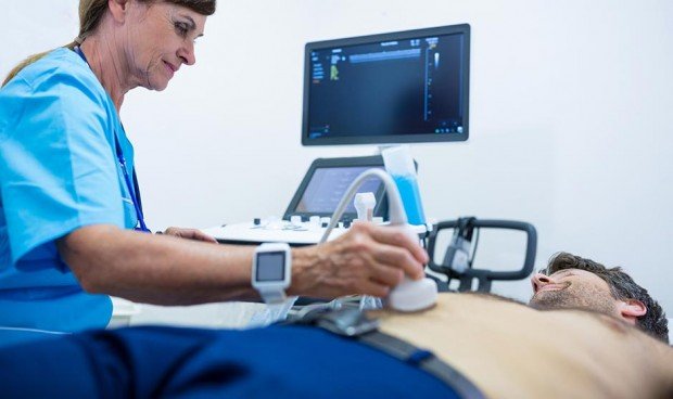
“There are more and more publications affecting the inappropriate use of Radiological tests that in many cases questionable justification is indicated, at the same time that the information that is passed on to the patient (by the attending physician and radiologist) is Insufficient “. With this hypothesis, the Spanish Society of Medical Radiology (CIRAM) In a document in which they specify and justify 62 practices who – which should not be done In certain positions by professionals.
“Before ordering a diagnostic test You must answer a series of basic questions, such as whether a file guide will modify patient management (clinical context), if necessary At the moment or if it can or should be postponed, or if the required test is less harmful to the patient and the person providing more information,” they state in the document “Dos and Don’ts for Prescribing Physicians, Radiologists, and Patients”.
Techniques that should not be done
Thus, this guide developed by SERAM aims to describe a series of Recommendations for radiological examinations that should not be performed, Directed to Prescribe physicians, radiologists, and patients. It should be promoted by radiology services, as good radiological practice, in cooperation and in agreement with the rest of the disciplines that order various imaging tests, for disease prevention, diagnosis and monitoring. These recommendations seek Reducing the use of old techniques of questionable efficacy and usefulness.
So this is all Techniques that society recommends not to implement, with explanations, corresponding details, and exceptions the radiologist may encounter.
-
Imaging tests (CT/MRI) for patients with symptoms suggestive of idiopathic primary headache
-
Plain X-rays in head injuries, except for the suspected non-accidental cause
-
Imaging tests for uncomplicated low back pain without warning signs
-
Imaging tests for uncomplicated neck pain without warning signs
-
Barium enema to evaluate colon diseases
-
Barium studies in inflammatory bowel disease
-
Routine chest x-rays before surgery
-
Follow-up with imaging techniques in benign solid pulmonary nodules
-
Abdominal computed tomography in pediatric patients with suspected acute appendicitis
-
Administration of intravenous contrast without prior safety examination
-
Daily chest x-rays for patients admitted to the Intensive Care Unit (ICU)
-
Chest X-ray after routine thoracentesis
-
Imaging tests to detect metastases in patients with breast cancer and breast cancer
-
Imaging tests to exclude metastases in patients who have had breast cancer surgery with curative intent and who are asymptomatic
-
Breast surgery for suspicious nodules without trying a percutaneous biopsy before
-
Early detection of breast MRI in patients without risk factors
-
Screening mammograms in women younger than 40 who do not have risk factors
-
Imaging techniques in patients with first episode of nontraumatic throat pain
-
Plain radiography routinely in ankle injuries
-
Conventional radiology studies to exclude bone metastases
-
Surgery as the primary treatment for osteosarcoma. An alternative to percutaneous techniques
-
Surgery as initial treatment for a patient with calcific tendinitis of the shoulder
-
Replace it with minimally invasive techniques
-
Ionizing radiation imaging techniques for assessing sacroiliitis activity. An alternative to an MRI
-
Approach central venous accesses without ultrasound guidance
-
Arteriography in the initial diagnosis of lower GI bleeding. An alternative to computed tomography angiography
-
Arteriography in the initial diagnosis and treatment planning of peripheral arterial disease
-
Plain radiography in suspected intussusception in pediatric patients
-
Pelvic X-ray for suspected hip dysplasia in children under 4 months of age
-
Routine imaging studies in children with acute uncomplicated bacterial sinusitis
-
Regular lateral radiography of the skull in children with obstructive sleep apnea syndrome (SHAS)
-
Neuroimaging studies in pediatric patients with primary headache
-
Barium studies in the diagnostic evaluation of children with inflammatory bowel disease (IBD)
-
X-ray of the pelvis in patients with multiple trauma undergoing computed tomography examination of the body
-
Abdominal X-ray in suspected acute diverticulitis
-
Computed tomography of patients with acute pancreatitis with unambiguous clinical presentation and elevated amylase and lipase
-
Intravenous urography (IVU) as a first choice in patients with acute flank pain and suspected renal colic
-
Abdominal X-ray in suspected acute pyelonephritis
-
Abdominal X-ray in acute abdomen, except for suspected obstruction or perforation of the intestine
Although it may contain statements, statements, or notes from health institutions or professionals, the information in medical writing is edited and prepared by journalists. We recommend that the reader be consulted on any health-related question with a healthcare professional.

“Social media evangelist. Student. Reader. Troublemaker. Typical introvert.”

:quality(85)/cloudfront-us-east-1.images.arcpublishing.com/infobae/TEQF6EONZRFGLLLDIDD4L2O4EE.jpg)

:quality(75)/cloudfront-us-east-1.images.arcpublishing.com/elcomercio/XU32LRAEZFDDPNVHLFU3CKVBYY.jpg)


More Stories
Venezuela ranks fourth in female leadership in science and technology in Latin America
In Portuguesa and Sucre they explore the wonderful world of science
The university court overturns the expulsion of two teachers and a chemical sciences student